Experimental Background Knowledge
Hemocytometer Overview
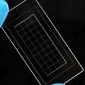
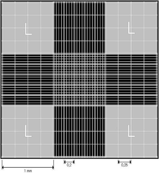
The Hemocytometer, specifically the model XB-K-25, is a precision instrument designed for counting cells. This device is divided into 25 distinct compartments, each serving as a critical area for accurate cell enumeration. The entire counting area of the Hemocytometer spans 1mm², providing a standardized platform for consistent measurements. Within this 1mm² area, each small square occupies an area of 1/400mm², ensuring that the cell counts are both precise and reproducible.
To better understand the structure and function of the Hemocytometer, consider the following breakdown:
| Feature | Description |
|---|---|
| Total Counting Area | 1mm² |
| Number of Compartments | 25 |
| Area of Each Square | 1/400mm² |
This design allows researchers to count cells with high accuracy, making it an essential tool in various biological and medical applications.
Counting Steps
Preparing the Cell Counting Plate
To ensure accurate cell counting, meticulous preparation of the Hemocytometer is crucial. Begin by thoroughly cleaning the counting plate with 75% ethanol to eliminate any potential contaminants. This step is essential as even minute residues can interfere with the accuracy of the count.
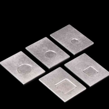
Next, carefully place the coverslip over the counting plate. The coverslip should be positioned with precision to ensure that it rests evenly on the grooves, creating a uniform chamber for the cell suspension. Any misalignment can lead to uneven distribution of cells, thereby skewing the count.
Once the coverslip is in place, transfer the counting plate to an ultraclean table. This environment is free from dust and other particulates that could compromise the integrity of the cell suspension. The ultraclean table provides a controlled setting where the subsequent steps of cell suspension preparation and counting can be performed with minimal risk of contamination.
By following these meticulous steps, you lay the groundwork for precise and reliable cell counts, ensuring that your experimental data is both accurate and reproducible.
Preparing the Cell Suspension
To prepare the cell suspension for counting, the first step involves digesting the cells to ensure they are in a suitable state for analysis. This process typically requires the use of enzymes or other reagents that break down the extracellular matrix, freeing the cells from their attachments. Once the cells are adequately digested, a precise volume of the suspension is extracted. It is common practice to take between 50 to 100 microliters of the cell suspension, ensuring that the sample is neither too dilute nor too concentrated, which could affect the accuracy of the count.
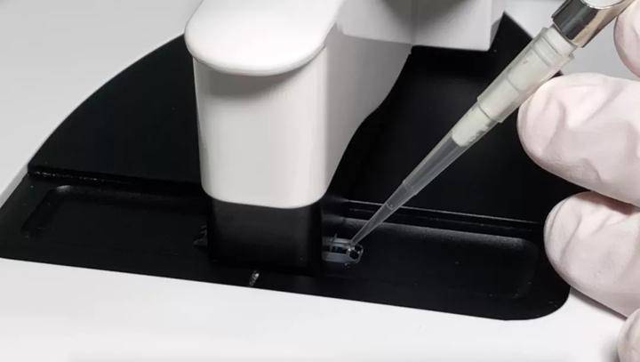
After obtaining the required volume, the suspension is carefully siphoned into the designated counting cells of the plankton counting chamber. This step demands precision and care to avoid introducing air bubbles or spilling the suspension, which could compromise the counting process. The hemocytometer, with its structured grid, is designed to facilitate accurate cell enumeration by providing a standardized area for counting.

In summary, the preparation of the cell suspension is a critical step that sets the stage for accurate cell counting. By carefully digesting the cells and precisely transferring the suspension, researchers ensure that the subsequent counting steps are based on a reliable and representative sample.
Cell Counting Plate under the Microscope
When examining the cell counting plate under a microscope, the structure becomes evident. The plate is meticulously divided into two distinct counting cells by an H-shaped groove, each containing 9 large squares. The central large square within each counting cell is further subdivided into 25 medium squares, creating a grid-like pattern that facilitates precise cell counting. This design ensures that cells can be accurately enumerated, providing crucial data for determining cell density and viability in various biological experiments.
Counting Methods
When performing cell counting using a Hemocytometer, two primary methods are employed: counting cells within large squares and within middle squares. Each method offers a distinct approach to accurately determine the density of cell suspensions.
Large Squares Method
In this method, the central large square, which is further divided into 25 medium squares, is used for counting. The formula for calculating the cell suspension density is as follows:
[ \text{Cell Density} = \frac{\text{Total Cells Counted}}{\text{Volume of Large Square}} ]
The volume of the large square is typically 1 mm³, making the calculation straightforward and precise.
Middle Squares Method
Alternatively, the middle squares method involves counting cells within the 25 medium squares that comprise the central large square. This method provides a more detailed count, especially useful for suspensions with varying cell densities. The formula for cell suspension density using this method is:
[ \text{Cell Density} = \frac{\text{Total Cells Counted in 25 Medium Squares}}{\text{Volume of 25 Medium Squares}} ]
The volume of each medium square is 1/25 mm³, ensuring a high level of accuracy in the cell density calculation.
By employing these methods, researchers can effectively determine the concentration of cells in a given suspension, facilitating accurate experimental results and reproducibility.
Cleaning the Cell Counting Plate
Proper cleaning of the cell counting plate is crucial to maintain its accuracy and longevity. After each use, the plate should be meticulously cleaned to remove any residual cell debris or contaminants. Begin by rinsing the counting plate with 75% ethanol, ensuring that all surfaces are thoroughly soaked. This step helps to disinfect the plate and eliminate any organic matter that could interfere with future cell counts.
Once the ethanol has been applied, it is essential to wipe the plate dry using a lint-free cloth or tissue. This prevents any remaining ethanol from pooling on the surface, which could potentially damage the delicate micro-structures of the counting grid. After drying, the plate should be stored in a clean, dust-free environment, typically within a dedicated storage box. This ensures that the plate remains protected from dust and other airborne particles when not in use.
It is important to note that while alcohol is an effective cleaning agent, it should not be used in excess. Avoid soaking the counting board in alcohol for prolonged periods, as this can lead to the degradation of the plate's materials over time. Instead, use a gentle, controlled approach to ensure that the plate is hygienically clean without causing unnecessary wear.
By following these cleaning protocols, you can ensure that your cell counting plate remains in optimal condition, providing reliable results for all your cell counting experiments.
Related Products
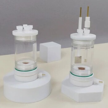
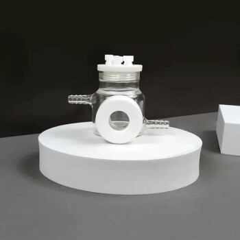
- Multifunctional Electrolytic Electrochemical Cell Water Bath Single Layer Double Layer
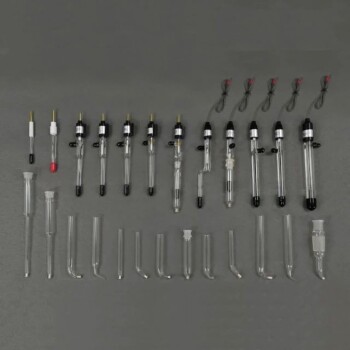
- Reference Electrode Calomel Silver Chloride Mercury Sulfate for Laboratory Use
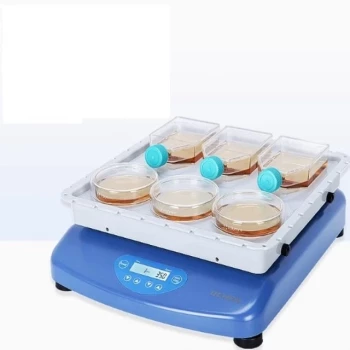
- Laboratory Oscillating Orbital Shaker
- Optical Water Bath Electrolytic Electrochemical Cell
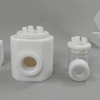
- Zooplankton Plankton Counting Chamber for Plankton Eggs and Ascaris Eggs
Related Articles
- The Thermodynamic Paradox: Balancing Precision and Safety in Electrolytic Cells
- The Thermodynamics of Consistency: Mastering the Invisible Variable in Electrolysis
- The Art of the Shutdown: Engineering Reliability in Electrochemical Cells
- The Architecture of Stability: Mastering Control with Double-Layer Electrolytic Cells
- The Silence of the Seal: Why Electrochemical Precision is a Battle Against the Atmosphere