The specific method utilized to analyze grinding-induced damage was Scanning Electron Microscopy (SEM). This imaging technique was applied directly to the ceramic material to characterize defects found on both the exterior and within the underlying material layers.
Core Takeaway: To fully understand the impact of the grinding process, researchers employed Scanning Electron Microscopy (SEM). This approach provided the necessary resolution to visually assess structural compromises in ceramics at both the surface and subsurface levels.
The Scope of the Analytical Approach
Evaluation of Ceramic Material
The analysis focused specifically on ceramic material. Because ceramics are inherently brittle and hard, standard visual inspections are often insufficient for detecting the micro-structural changes caused by mechanical processing.
SEM provides the high magnification required to observe the specific fracture mechanics and material removal mechanisms typical in ceramics.
Dual-Layer Investigation
The study did not limit its scope to the visible exterior. The SEM analysis was structured to capture two distinct categories of structural failure:
- Surface Damages: Identifying direct topographical defects, scratches, and voids created by the grinding wheel interface.
- Subsurface Damages: Examining the material layers beneath the surface to identify deep cracks or structural alterations that are not visible to the naked eye.
Understanding the Analytical Constraints
Qualitative vs. Quantitative Data
While SEM is exceptional for visualization, it primarily provides qualitative morphological data. It reveals what the damage looks like (e.g., crack propagation or pulverization) but does not inherently measure the residual stress or mechanical strength reduction caused by that damage without auxiliary testing.
The Challenge of Subsurface Imaging
Analyzing subsurface damage via SEM usually requires specific sample preparation, such as cross-sectioning.
If the cross-sectioning is not performed precisely, it can be difficult to distinguish between damage caused by the original grinding process and damage introduced during the preparation of the sample itself.
Implications for Material Assessment
When reviewing the results of this analysis, consider your specific evaluation goals:
- If your primary focus is cosmetic quality: The SEM analysis of surface damage will reveal the roughness and finish consistency of the ceramic.
- If your primary focus is mechanical reliability: Pay closest attention to the subsurface damage findings, as hidden micro-cracks are often the initiation points for catastrophic component failure.
By utilizing SEM, the analysis bridges the gap between visible surface flaws and critical internal structural integrity.
Summary Table:
| Feature | Surface Damage Analysis | Subsurface Damage Analysis |
|---|---|---|
| Focus Area | Topographical defects & scratches | Hidden micro-cracks & structural flaws |
| Key Insight | Aesthetic finish & roughness | Mechanical reliability & failure points |
| Detection Method | Direct SEM imaging | Cross-sectioning + SEM imaging |
| Material Impact | Surface voids and tool marks | Initiation points for catastrophic failure |
Elevate Your Material Analysis with KINTEK
Precision grinding requires precision analysis. At KINTEK, we understand that the integrity of your ceramic components depends on identifying both visible and hidden defects. Whether you are processing brittle materials or developing advanced composites, our comprehensive range of laboratory equipment—from crushing and milling systems to high-temperature furnaces and hydraulic presses—is designed to meet the most rigorous research standards.
Don't let subsurface micro-cracks compromise your mechanical reliability. Contact KINTEK today to discover how our high-performance tools and consumables can optimize your material characterization and production workflows. Let us help you achieve superior structural integrity.
Related Products
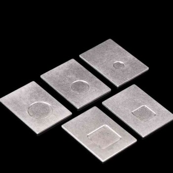
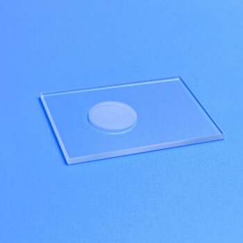
- Customizable XRD Sample Holders for Diverse Research Applications
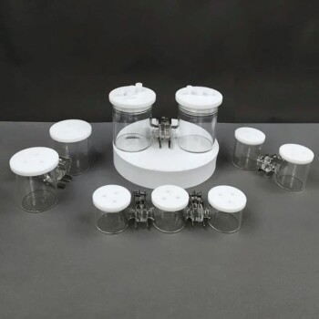
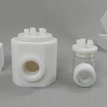
- H Type Electrolytic Cell Triple Electrochemical Cell
- XRD Sample Holder X-ray Diffractometer Powder Slide
- Side Window Optical Electrolytic Electrochemical Cell
People Also Ask
- What are the applicable sample dimensions for the sample holder? Ensure a Perfect Fit for Your Lab Samples
- What is the typical function of the sample holder in an electrochemical experiment? It's the Active Working Electrode
- How should a suitable sample be selected and secured in the holder before an experiment? Ensure Unshakeable Stability for Reliable Data
- How should a sample holder be cleaned and inspected before use? Ensure Reliable Lab Results
- What are the specific storage requirements for a sample holder? Protect Your Lab's Critical Assets