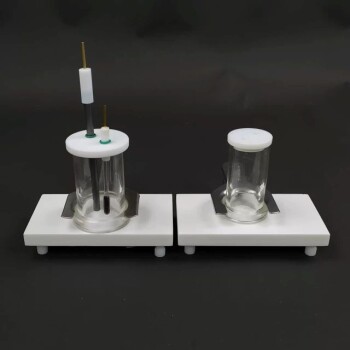
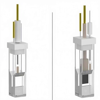
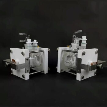
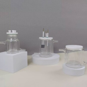
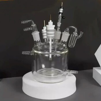
Precise control of the electrolyte layer thickness is the defining requirement for a transmission-type in-situ XAFS cell, with the optimal depth typically maintained at approximately 1.5 mm. This specific dimension is engineered to minimize X-ray photon absorption by the liquid medium while simultaneously preserving a fully functional three-electrode electrochemical environment.
The core challenge in cell design is balancing optical transparency with chemical functionality. The 1.5 mm thin-layer architecture is the critical standard that prevents the electrolyte from masking the signal, ensuring high-quality data on the catalyst’s oxidation states.
Optimizing for X-ray Transmission
The 1.5 mm Thickness Standard
To acquire usable spectroscopic data, the design must strictly limit the path length of the X-ray beam through the liquid.
Research indicates that maintaining an electrolyte layer of approximately 1.5 mm is the ideal specification. This dimension is not arbitrary; it represents a calculated effort to reduce the physical volume of liquid the beam must penetrate.
Minimizing Photon Absorption
The primary adversary in transmission-type XAFS is the absorption of X-ray photons by the electrolyte itself.
If the liquid layer exceeds the 1.5 mm threshold, the electrolyte absorbs a significant portion of the incident X-rays before they interact with the catalyst. By enforcing a thin-layer design, the cell ensures that enough photons reach the detector to generate a clear, analyzable signal.
Maintaining Electrochemical Fidelity
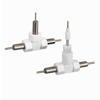
The Three-Electrode Requirement
Despite the geometric constraints required for spectroscopy, the device must operate as a legitimate electrochemical cell.
The design must accommodate a functional three-electrode setup within the confined space. This ensures that the potential control and current measurement remain accurate, allowing the researcher to drive the reaction exactly as they would in a standard reactor.
Capturing Dynamic Reaction Data
The ultimate goal of this precise design is to enable the collection of high-quality absorption spectra, such as Copper K-edge spectra.
By maintaining the 1.5 mm geometry, researchers can effectively monitor real-time changes during the reaction process. This clarity is essential for determining the oxidation states and coordination environments of the catalyst as they evolve.
Understanding the Trade-offs
Signal Intensity vs. Electrochemical Stability
Designing these cells involves an inherent compromise between the needs of the physicist (X-ray transmission) and the chemist (reaction stability).
The Risks of Improper Sizing
If the cell design ignores the 1.5 mm constraint in favor of a larger electrolyte volume, the resulting X-ray absorption by the liquid will degrade the signal-to-noise ratio, rendering the spectra unusable.
Conversely, if the cell is made too thin in an attempt to maximize transmission, it may become difficult to house the three-electrode system or maintain stable electrochemical conditions. The 1.5 mm specification acts as the critical "middle ground" where both physics and chemistry function correctly.
Making the Right Choice for Your Goal
When specifying or building an in-situ XAFS cell, prioritize the internal geometry above all other features.
- If your primary focus is Signal Quality: Strictly adhere to the 1.5 mm electrolyte thickness to minimize photon loss and ensure high-fidelity spectral data.
- If your primary focus is Reaction Mechanism Analysis: Ensure the thin-layer design still supports a robust three-electrode configuration to accurately correlate oxidation states with applied potentials.
The most effective cell design is one that treats the electrolyte thickness not as a variable, but as a fixed optical component of the spectroscopic system.
Summary Table:
| Design Feature | Specification | Impact on Research |
|---|---|---|
| Electrolyte Layer Thickness | ~1.5 mm | Minimizes X-ray photon absorption while maintaining liquid flow. |
| Electrode Configuration | Three-electrode system | Ensures accurate potential control and reaction driving. |
| Optical Goal | Transmission Transparency | Maximizes signal-to-noise ratio for K-edge spectra. |
| Chemical Goal | Electrochemical Fidelity | Correlates oxidation states with real-time reaction data. |
Elevate Your In-Situ Research with KINTEK Precision
Unlock the full potential of your spectroscopic analysis with KINTEK’s specialized laboratory solutions. Whether you are studying catalyst oxidation states or coordination environments, our high-performance electrolytic cells and electrodes are designed to meet the rigorous 1.5 mm geometry standards required for flawless XAFS data.
From high-temperature furnaces for material synthesis to advanced electrochemical research tools, KINTEK provides the reliability and precision your lab demands. Don't let poor cell design compromise your signal quality.
Contact our technical experts today to find the perfect fit for your research goals!
References
- Shikai Liu, Qian He. Alkali cation-induced cathodic corrosion in Cu electrocatalysts. DOI: 10.1038/s41467-024-49492-7
This article is also based on technical information from Kintek Solution Knowledge Base .
Related Products
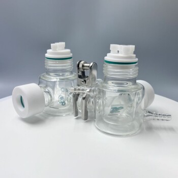
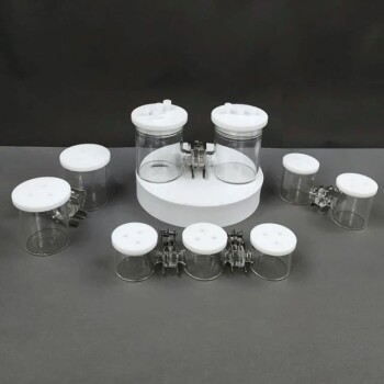
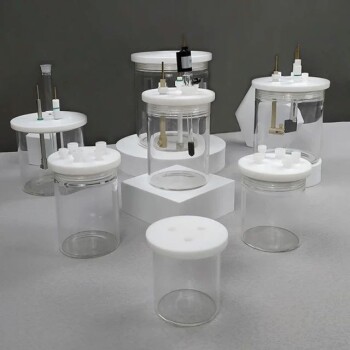
- H-Type Double-Layer Optical Electrolytic Electrochemical Cell with Water Bath
- H Type Electrolytic Cell Triple Electrochemical Cell
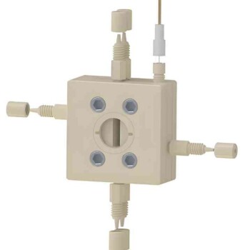
- Electrolytic Electrochemical Cell with Five-Port
- Side Window Optical Electrolytic Electrochemical Cell
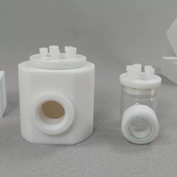
- PTFE Electrolytic Cell Electrochemical Cell Corrosion-Resistant Sealed and Non-Sealed
People Also Ask
- How is the electrolyte managed in H-type electrolytic cells for specific reactions? Achieve Precise Control and High Purity
- What checks should be performed before using an H-type electrolytic cell? Ensure Experiment Safety and Data Accuracy
- What is important regarding temperature control for the H-type electrolytic cell? Ensure Precision and Data Integrity
- What checks should be performed on the H-type electrolytic cell before use? Ensure Accurate Electrochemical Data
- What is the purpose of the double-layer structure in the H-type electrolytic cell? Achieve Precise Thermal Control