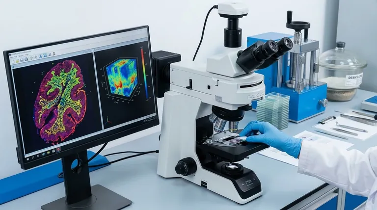
For infrared (IR) imaging of biological samples, the most widely utilized and powerful technique is Fourier Transform Infrared (FTIR) Microspectroscopy. This method combines a standard IR spectrometer with a microscope, allowing you to generate chemically-specific images that reveal the spatial distribution of key biomolecules like proteins, lipids, and nucleic acids within a tissue section or group of cells.
The core challenge of IR imaging in biology is not just choosing a technique, but managing the overwhelming IR signal from water, which can obscure the molecular data you seek. Therefore, your choice of both the instrument and the sample preparation method is critical for success.
What is Infrared Imaging? A Chemical Map
Infrared imaging, also known as vibrational microspectroscopy, is fundamentally different from standard optical microscopy. Instead of just visualizing morphology, it provides information about the chemical composition of the sample.
Beyond a Picture: Creating a Hyperspectral Image
An IR microscope measures a full infrared spectrum at every single pixel in the image. This creates a "hyperspectral data cube," which is a stack of images where each layer corresponds to the absorption of light at a specific IR frequency.
By analyzing this data, you can generate false-color images that map the concentration and distribution of specific chemical components across your sample.
The "Fingerprint" Region: Identifying Key Molecules
The mid-infrared region of the spectrum (approximately 4000-400 cm⁻¹) causes molecules to vibrate. Different chemical bonds (like C=O in proteins, C-H in lipids) vibrate at characteristic frequencies.
The region from roughly 1800 to 900 cm⁻¹ is known as the "fingerprint region" because it contains a complex pattern of peaks that is unique to a specific molecule. By analyzing this region, you can identify and quantify the main classes of biomolecules.
The Dominant Technique: FTIR Microspectroscopy
While other methods exist, FTIR microspectroscopy is the workhorse of the field for its balance of sensitivity, speed, and versatility.
Why FTIR? Speed and Sensitivity
Modern Fourier Transform Infrared (FTIR) instruments collect all frequencies of light simultaneously, a significant advantage over older methods. This results in a much higher signal-to-noise ratio and dramatically faster acquisition times, which are essential for mapping large areas of a biological sample.
The "Micro" Advantage: Spatial Resolution
Pairing the FTIR spectrometer with a microscope allows you to focus the IR beam down to a small spot. By raster-scanning this beam across the sample or using a focal plane array (FPA) detector, you can build the hyperspectral image pixel by pixel, resolving features on the scale of tens of microns down to a few microns.
The Core Challenge: Overcoming Water Interference
The single greatest obstacle in IR analysis of biological samples is water.
Why Water is a Problem
Liquid H₂O has extremely strong and broad absorption bands in the mid-IR range, particularly around 1640 cm⁻¹. This signal is so intense that it can completely saturate the detector and mask the crucial Amide I band of proteins, which is essential for studying protein structure and concentration.
Solution 1: Sample Drying and Fixation
The most common approach is to remove the water. Biological tissues are typically sectioned using a microtome, placed on a special IR-transparent slide (like CaF₂ or BaF₂), and then dried.
This can be done by air-drying, freeze-drying (lyophilization), or using chemical fixatives like formalin or ethanol, similar to standard histology. This effectively eliminates the water signal, providing clean, high-quality spectra of the remaining biomolecules.
Solution 2: Isotopic Exchange with Heavy Water (D₂O)
For studying samples in a more "native" or hydrated state, such as live cells, the H₂O can be exchanged with deuterium oxide (D₂O), or "heavy water."
The O-D bond in D₂O absorbs at a much lower frequency (around 1210 cm⁻¹), shifting the massive water peak out of the way and revealing the protein, lipid, and nucleic acid signals in the fingerprint region.
Understanding the Trade-offs: Measurement Modes
How the IR light interacts with your sample is another critical choice, with each mode offering distinct advantages.
Transmission
In transmission mode, the IR beam passes directly through a very thin sample. This mode generally provides the highest quality, most quantifiable spectra but requires meticulously prepared, thin tissue sections (typically 5-10 µm).
Reflection (Transflection)
More commonly, samples are analyzed in transflection mode. The tissue is placed on a reflective slide (like a mirrored or Low-e slide). The IR beam passes through the sample, reflects off the slide's surface, and passes back through the sample to the detector. It is more convenient but can sometimes introduce spectral artifacts.
Attenuated Total Reflectance (ATR)
ATR-FTIR imaging is a powerful surface-sensitive technique. The sample is brought into firm contact with a high-refractive-index crystal (like germanium). The IR light does not pass through the sample; instead, an "evanescent wave" penetrates only a few microns into the sample's surface.
This is excellent for getting high-quality spectra from the surface of thick or highly absorbing samples without any preparation. Its short pathlength naturally minimizes water interference, making it a strong choice for analyzing hydrated samples.
Emerging Frontiers in IR Bio-Imaging
The field is constantly evolving with new technologies that push the boundaries of speed and resolution.
Synchrotron IR: For Ultimate Resolution
Using a synchrotron light source provides an IR beam that is up to 1000 times brighter than a conventional thermal source. This allows for diffraction-limited spatial resolution, enabling the chemical imaging of single cells and even subcellular organelles.
Quantum Cascade Lasers (QCLs): For Unprecedented Speed
Instead of a broad thermal source, these systems use high-power, tunable lasers. While they don't typically collect the full spectrum, they can be tuned to a few key frequencies to map specific molecules (like total protein or lipid) across very large areas in a matter of minutes, rather than hours. This is transforming the potential for high-throughput clinical applications.
Making the Right Choice for Your Goal
Your selection of technique and sample preparation depends entirely on your research question.
- If your primary focus is diagnostic histopathology: Use FTIR microspectroscopy in transmission or transflection mode on thin, dried, and fixed tissue sections to identify biochemical markers of disease.
- If your primary focus is studying live cells or dynamic processes: Consider ATR-FTIR imaging or work in a sealed liquid cell after exchanging the media with D₂O to maintain a hydrated environment.
- If your primary focus is subcellular chemical analysis: You will likely require the high brightness and spatial resolution afforded by a Synchrotron-IR source.
- If your primary focus is high-throughput screening of many samples: QCL-based imaging offers the speed required to rapidly map the distribution of a few key biomarkers.
Ultimately, mastering infrared imaging of biological samples is about controlling your variables to isolate the molecular signals that matter most.
Summary Table:
| Technique | Key Advantage | Best For |
|---|---|---|
| FTIR Microspectroscopy | High sensitivity & speed | General chemical mapping of tissues |
| ATR-FTIR Imaging | Minimal sample prep, surface-sensitive | Hydrated samples, live cells |
| Synchrotron-IR | Ultimate spatial resolution | Subcellular analysis |
| QCL Imaging | Unprecedented speed | High-throughput screening |
Ready to unlock the full potential of IR imaging in your research? KINTEK specializes in providing advanced lab equipment and consumables for all your FTIR and biological imaging needs. Whether you're working with tissue sections, live cells, or require high-throughput solutions, our experts can help you select the right tools to achieve precise, chemically-specific results. Contact us today to discuss how we can support your laboratory's success!
Visual Guide

Related Products
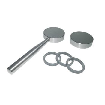
- XRF & KBR plastic ring lab Powder Pellet Pressing Mold for FTIR
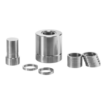
- XRF & KBR steel ring lab Powder Pellet Pressing Mold for FTIR
People Also Ask
- How are samples prepared for XRF analysis? Achieve Accurate and Reliable Results
- How does the selection of a pressure mold affect the performance of all-solid-state batteries? Expert Pelletizing Guide
- What is the XRF method of sampling? Achieve Accurate Elemental Analysis with Proper Sample Prep
- What is the XRF pressed pellet method? A Fast, Cost-Effective Sample Prep Guide
- What is the purpose of using a mold for pellet pressing when preparing catalyst test samples? Ensure Data Consistency